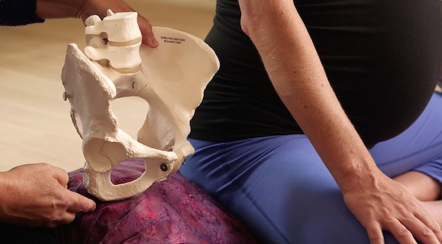
The three levels of the pelvis are discussed in the Spinning Babies® Workshop as they relate to baby’s journey through them. In the body balancing portion of the workshop, we discuss the muscles and soft tissues. I thought you’d like a little tour of the pelvis as these structures are dehttps://www.spinningbabies.com/wp-content/uploads/2019/10/sample3-1.pngd together.
At the Top of the Pelvis

The first pelvic opening is the inlet, or brim. There are three joints which work a bit like hinges but have the strong support of muscles and ligaments to keep these joints stable. The two joints in the back are the sacroiliac joints, or SI joints. One or both are often tender, alerting us to add balance and rewarding us with comfort when we do. The joint in the front is the symphysis pubis. We nickname the thick bony portions protecting the cartilage as “the pubic bone.” This joint doesn’t move much, or isn’t supposed to, because when it does it can hurt. Two round ligaments wrap over the outer edges of the pubic bone. They come from the uterus (which is from above if in mid or late pregnancy) and end beneath the pubic bone in the connective tissues. They help hold the uterus to the pelvis in front, but if one or both are tight, they “zing” when touched. The broad ligament wraps the uterus from the sides, covering much of the front sides of the uterus. If these are tight, baby’s kicks can actually hurt. It can even feel as if baby is kicking the ribs, especially on the right side.

The muscles supporting the top of the pelvis include two guiding branches of the psoas (so-as) which cross from the back to the front and drape the leg joints on their way to anchor to the upper thigh (attaching to the lesser trochanters). The psoas can guide the baby into the pelvis, or hold the baby up if they’ve shortened their length due to sitting with knees higher than the level of the hips, or not walking regularly throughout the day. The abdominal muscle layers support (ideally) the front, and muscles from the spine to the back wings of the pelvis also play a part in helping the pelvis and torso be stable while yet allowing movement.
When these muscles, joints, and ligaments are supple and supportive, they guide baby to “engage,” or settle down, into the pelvic brim near birth time.
Halfway Down the Pelvis

In the midpelvis, the pelvic floor acts like a sling to support the organs above it, including the bladder, rectum, and uterus. A long, supple pelvic floor moves with breathing, matching the respiratory diaphragm by coming up when the breath comes in and going down a little distance when the breath goes out. An opening in the pelvic floor not only allows baby to pass through but turns baby’s head to the diagonal (called the oblique diameter in anatomy textbooks) so baby fits through the midpelvis opening. Making the pelvic floor longer with the squats we demonstrate in class means a more flexible and responsive opening to reduce pain and allow head rotation better than if the pelvic floor is short and tight.
Sidelying Release, one of the Three Sisters of Balance soothes and lengthens the pelvic floor for at least a few hours. When baby is midway down the pelvis, give it a try! Sidelying Release helps the muscles to the sacrum and tailbone, helping both the midpelvis and outlet have space. These muscles include the piriformis and its gang called The Deep Six, the glutes which cover the Six, and the hamstrings at the back of the leg. Muscles on the thighs which rotate the leg at the leg socket sometimes hurt in labor. Giving them a good range of motion in pregnancy with the Daily Essentials, and giving them flexibility with The Three Sisters of Balance may allow ease at the outlet as well as all the way up the pelvis.
At the Bottom of the Pelvis

In the outlet, the last opening of the pelvis, baby turns again to face directly back, towards your tailbone and sacrum. The mobility of the sacrum allows it to move outward in late dilation. In addition to the sacrum having moved outward, the mobility of the joint connecting the coccyx to the sacrum assures maximum space in the posterior portion of the pelvis.

The location of the sacrum in relation to the front of the pelvis, specifically the distance of the sacrum to the ischial tuberosities. The distance here is increased with the sacrotuberous ligament release that I dehttps://www.spinningbabies.com/wp-content/uploads/2019/10/sample3-1.png on the blog. The mobility of the sacrum is helped in part with calf stretches, anterior pelvic tilts, and squats. The flexibility of the sacrum is enhanced with the standing sacral release. These techniques are in our techniques section.
Enjoy this post? You also might like:
- Maternal Positions at the Levels of the Pelvis
- Birth-Related Anatomy
- Three Levels to Avoiding a Cesarean
- The Difference a Sidelying Release Makes
Upcoming Workshops
[tribe_events_list limit=”4″]